Premium
Vet on call: Dealing with rumen flukes attack in goats

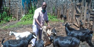
Gerishon Kamotho feeds his goats in Elburgon, Nakuru County. Animals, including goats, should be dewormed every three months to break the life-cycle of parasites.
What you need to know:
- To have an adequate, efficient and effective national animal health service, paravets should work under specified veterinary doctors so that the seniors can offer expertise where case complexity exceeds the training of the paravets.
- Paramphistomes have a complicated life cycle. The adult worms lay eggs that are expelled with faeces. The eggs hatch in water and the tiny hatchlings called miracidia, actively seek a suitable snail host.
- As they approach maturity, the paramphistomes migrate back into the rumen where they attach to the wall, mature and start laying eggs.
- Animals should be dewormed every three months to break the parasite life cycle. Snails should be eliminated from pastures and animals prevented from grazing in marshy pastures.
I worked with June for the first time last week when she wrote me an email requesting for assistance. She is a paravet running her private practice in Kirinyaga.
To have an adequate, efficient and effective national animal health service, paravets should work under specified veterinary doctors so that the seniors can offer expertise where case complexity exceeds the training of the paravets.
I am always happy and willing to work with paravets from any part of the country whenever they call for assistance.
June had two cases, the first one was of goats dying of pneumonia-like disease that spread fast in the herd.
She had sent a carcass to the laboratory and contagious caprine pleuropneumonia (CCPP) was diagnosed.
She wanted to know how she could treat the remaining sick goats and prevent the healthy ones from getting infected.
I told her she had a very difficult-to-deal-with disease on her hands. I advised the medicines to give but cautioned she could still lose many of the sick animals.
She would also cover the healthy goats with one of the medicines specific for the CCPP bacteria. The surviving goats would be vaccinated against the disease at least five days from the date of the last injection.
June’s second case was dystocia, which means a difficult birth in a goat. She said the foetus had failed to enter the birth canal since the cervix had not opened wide enough.
June could therefore not extract the kid from the uterus. She advised the owner to slaughter the goat. If the goat was of high value, June could have referred the case to a veterinary surgeon for a caesarean surgery.
Every part of the goat carcass looked normal until June opened the large stomach called the rumen. A greater part of the inner lining was unusual.
It looked like it had ovoid pink berries attached to the lining. The normal appearance is that of a green towel.
June told me the farmer looked at the unsightly rumen appearance and said the whole goat carcass should be disposed of. June wanted to know what the condition was and if the meat was fit for human consumption.
A complicated life-cycle
I examined the pictures she sent me on WhatsApp and just smiled. What she was seeing was common in sheep, goats, cattle and buffaloes that feed in water-logged areas or where there is stagnant water.
I advised the rumen could be trimmed in the areas affected but the unaffected parts and the rest of the carcass was good for human consumption.
The goat owner rejected the advice and said he would boil the whole rumen for his dogs. That was fine with me. You see, it is not always about the safety of meat.
The appearance must also be pleasing to the eye to give what is called aesthetic satisfaction. The condition was a case of paramphistomosis or infestation by rumen flukes known as paramphistomes (Paramphistomum cervi).
The parasites appeared pink because of the blood they had sucked from the rumen blood vessels. The adult worms usually do not cause obvious disease in adult animals.
However, excessive blood loss may cause weakness especially in animals with other diseases or pregnant ones. This could have been the reason why the goat got dystocia.
Paramphistomes have a complicated life cycle. The adult worms lay eggs that are expelled with faeces. The eggs hatch in water and the tiny hatchlings called miracidia, actively seek a suitable snail host.
They dig into the snail where they transform into forms called cercariae. These later change into metacercariae and break out of the snail. They form cysts embedded on plants, such as grass, and wait to be consumed by livestock.
Once ingested, the cysts open up in the small intestines as immature paramphistomes. The parasites attach on the intestinal wall where they suck blood and cause tissue death.
The victim animal may develop diarrhoea with or without blood, dehydration and intestinal bacterial infection. Young animals tend to die in large numbers if the parasite load is heavy.
As they approach maturity, the paramphistomes migrate back into the rumen where they attach to the wall, mature and start laying eggs.
It is not known how long they can live in the rumen. There are dewormers that are capable of killing the parasites.
Animals should be dewormed every three months to break the parasite life cycle. Snails should be eliminated from pastures and animals prevented from grazing in marshy pastures.


