Dairy cows at a farm in Kirinyaga County.

Farmers have asked me many times whether it is possible for a livestock farm to be completely free of diseases. My outright answer, even straight from a deep midnight sleep is a resounding “no”. However, farms, regions and nations can be free of specific diseases.
Just like we cannot totally eradicate all diseases in human beings, we cannot do the same in livestock for various reasons. One major reason is that there are very many disease-causing organisms or pathogens in any environment. The level and method of interaction between the pathogens and livestock determines whether the animals will remain healthy or will get sick.
The second is that all animals carry microorganisms on their skin, in the fir and in their bodies throughout their life. Some of the microorganisms are infective, others harmless and others useful to the extent that they help the animals fight infections.
The infective organisms will cause disease when the animal’s environment changes and encourages their increase in numbers or production of toxic chemicals that then cause disease in the animal. A good example are the Clostridium perfrigenes bacteria in the intestines. When the diet suddenly has too much highly digestible sugars, the bacteria multiply heavily, produce toxins and cause severe sickness and death in both humans and animals.
A third reason we cannot completely eradicate diseases is that pathogens can be transported from one area to another through various methods including feeds, water, air, other materials and infected animals. This is why we always insist on good hygiene, biosecurity, quarantine of new animals introduced on the farm, vaccination of animals and prompt disease detection and treatment.
Fourth, new pathogens or modified ones keep emerging all the time. Others change themselves and resist death from commonly used medicines.
Finally, there are diseases that are non-infectious such as those caused by body dysfunction, injury or old age degeneration. Milk fever in cattle and post-calving ketosis are common non-infectious diseases in dairy cattle caused by body dysfunction.
Milk fever is caused by a fault in the body’s regulation of calcium between the blood and the deposits in the bones. Ketosis occurs when a high yielding cow gets insufficient carbohydrates during periods of high energy demand and is forced to burn its fat to generate energy. This is mainly during late pregnancy and early lactation.
Both diseases can be successfully treated if detected early enough before the body cells get irreparably damaged. Not withstanding good treatment, some animals may still fail to recover.
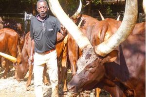
This week’s article is motivated by the latest feedlot project I have reported on in my recent articles. We have had additional disease challenges but nothing out of the ordinary. This week I visited the farm and found the animals have put on a lot of weight and have also changed body colour. All of them now have shiny coats showing their true breed colours. The animals are weighed once per week to monitor body weight changes.
The feedlot has not yet attained the target daily weight gain of 0.7 to 1.7kg per animal but the bulls are steadily progressing towards it. In a feedlot, one has to keep a close eye on the difference between the current body weight and the entry body weight. The difference between the two gives the overall weight gain.
Once the figure is divided by the number of days the animals have been on the farm, one gets the daily weight gain. Feedlot farming is like financial management. One has to deal a lot with figures, understand what they mean and use them to make decisions that are critical to the success and profitability of the business.
I was happy to note that the animals have now fully settled in the feedlot and their new diet. They are actually loving it a lot. Once they have fed, they cluster together with most lying down to digest the feed and add weight.
On my visit, there were new disease challenges although in only three cattle. One cow I had treated for mange had recovered from the disease but the same area around the left eye had now developed fungal and papilloma virus infections.
The finding was not surprising because the pathogens for the two diseases are always there in the environment and may also be on the animal’s skin. The mange infestation may have triggered the other two diseases by breaking the skin and providing the opportunity for infection.
There was more ringworm or fungal infection around the eye of the animal than the papillomas. The papillomas may heal on their own with time. I therefore prescribed iodine treatment by application on the infection patches twice daily till recovery. I observed the cow I prescribed iodine in the last visit, one week earlier, for the same problem was recovering well.
Another cow had a swollen lower rear leg up to the dew claws. I noticed the external lymph nodes were swollen but he cow was eating well. I concluded it was another case of mild lumpy skin disease or LSD. I had seen one in the last visit but it only had drying pinpoint nodules on the skin.
I was grateful that I had vaccinated the animals against the disease immediately they had come in. LSD is a viral disease that can be very devastating to cattle when fully expressed. It causes high fever, large round painful nodules in the skin and mucous membranes in unvaccinated animals.
For animals with appreciable immunity, the disease causes painless swelling of one or more limbs and the external lymph nodes. The nodules, in unvaccinated animals, may occur in the pulmonary and gastrointestinal systems. Such cases may not survive the disease.
Finally there was a bull which would limp on the left rear leg after standing from a sleeping position. I observed the animal and saw it was limping with the middle part of the leg just below the knee.
I examined the animal physically and found that the area below the knee had a healed injury possibly from a broken leg when the animal was young.
The observed lameness is characteristic in such cases. The limping stops after the animal stands up and walks around because the tissues with the old injury get resupplied with blood and oxygen. No treatment is given for the condition as it is temporary and mild.